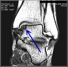
Is there chronic ankle pain after a bad sprain?
TALAR
DOME INJURY
Is there chronic ankle pain after a bad sprain? It could be a chip in the joint. These fractures usually occur as a result of an ankle injury or an ankle
sprain. Sadly, this injury is not usually appreciated on plain x rays, because cartilage usually appear as black space. There is not an actual space between the joint. The dome of the talus articulates with the tibia and fibula, and has a key role in ankle motion and in supporting your body weight when walking or standing. The dome is made up of bone (osteo) that is covered by a layer of articular cartilage (chondral). Articular cartilage is a smooth shiny material that allows the ankle bones to slide easily over each other as the ankle moves. When a part of the dome cracks or breaks off, it is referred to as an osteochondral fracture of the talar dome.
Fractures of the talar dome are generally
the result of inversion injuries of the ankle. They are located medially or
laterally with equal frequency and occasionally through both. Lateral talar
dome fractures are almost always associated with trauma, while medial talar
dome lesions can be traumatic or atraumatic in origin. Although the etiology in
atraumatic lesions is unclear, osteochondral fragments can separate from the
surrounding cartilage surface and dissect into the joint space. As these
osteochondral fragments become loose in the joint, they can cause pain,
locking, crepitus, and swelling.
DIAGNOSIS
 Clinical diagnosis of talar dome fractures
can be highly challenging and are often missed. The patient may have sustained
a fall or a twisting injury to the ankle and may generally ambulate with a
limb. In the acute setting, the symptoms of a talar dome fracture are similar
to and often occur with an ankle sprain. In lateral talar dome lesions,
tenderness is generally found anterior to the lateral malleoli, along the anterior
lateral border of the talus. In medial talar dome lesions, tenderness is
usually located posterior to the medial malleolus, along the posterior medial
border of the talar dome.
Chronic talar dome lesions, traumatic and
atraumatic osteochondritis dissecans lesions may have a clinical presentation
similar to that of arthritis. Typical findings include crepitus, stiffness,
and recurrent swelling with activity.
Diagnosis of talar dome lesions can often
be made with standard anteroposterior (AP), lateral, and mortise ankle
radiographs. However, repeated radiographs may be necessary because initial
films may appear normal. Secondary changes in the subchondral bone (visible on
plain radiographs) caused by a compression fracture of the articular
osteochondral surface may take weeks to appear. In addition, small chondral
fragments are radiolucent and not evident on standard radiographs.
Generally, the AP ankle view is best for
visualizing deep, cup-shaped medial lesions, although the lesions are often
appreciated on the mortise view as well. Lateral lesions are best visualized on
a mortise view and are generally thin and wafer-shaped. If suggested by the
clinical scenario, fractures not visualized with plain radiographs may require
magnetic resonance imaging (MRI) or computed tomography (CT).
TREATMENT
These lesions require treatment because of the high functional demands of the talar dome and the potential for
complications. Stage I, II, and III medial lesions can usually be treated
nonsurgically with six weeks in a nonweight-bearing cast. Adequate reduction
and immobilization are crucial for fracture healing and to avoid avascular
necrosis of the fracture fragment. Patients with stage III lateral lesions,
stage IV lesions, and persistent symptoms are generally treated surgically.
Treatment options for fragment excision range from arthroscopy with or without
subchondral bone drilling and graphing to open reduction internal fixation.